


The disease caused by a violation of the structure of the vascular walls, the thinning of which with pronounced stagnant processes of blood flow is called varicose veins. The disease often affects the lower extremities, although it can be localized in the rest of the body. According to the international classification of diseases of the tenth revision of the ICD 10, varicose veins are assigned the code 183, which includes four headings that describe various manifestations of the disease.
How did varicose veins develop?
The first mentions of varicose veins were found in ancient Greek papyri. History and confirmed scientific facts state that varicose veins of the lower extremities were found in the found Egyptian mummy - it can be argued that the disease has accompanied humanity throughout its existence.
Excellent doctors - Avicenna, Hippocrates, Galen tried to treat varicose veins of the lower extremities. In the 19th century, painful and traumatic treatments were used which consisted of dissecting the tissues of the thigh and lower leg to damage the saphenous veins, followed by a bandage. It was understood that in this way stagnant blood flow processes can be prevented and varicose veins can be eliminated. However, the methods left terrible, widespread scars on patients' bodies and contributed to damage to nerves, arteries, and disorders of lymphatic flow.
A little later, the history of the treatment of varicose veins received a positive breakthrough - in 1908 a metal probe was first used as a means of minimally invasive effect on the walls of blood vessels. The discovery of radiography allowed more accurate and efficient surgical interventions to eradicate varicose veins. Now when proper diagnosis and treatment of the disease is needed, duplex and triplex scans, powerful drugs, laser therapy, and sclerotherapy are used. Surgical intervention is used only in cases when varicose veins cannot be sparingly eliminated.
The main causes of the disease
Varicose veins are a great danger, the pathology has become "younger" - in the past it was mainly the elderly who suffered, now varicose veins are diagnosed in young patients, extremely rarely in children.
Causes of the disease:
- Genetic predisposition.
- Overweight, overweight, obesity.
- Sedentary inactive lifestyle.
- Bad diet, poor blood quality.
- Concomitant disease of the cardiovascular system.
- Professional activity.
- Prolonged standing, heavy physical exertion.
- Pregnancy and hormonal changes.
- Individual features of the structure of the vascular system.
- Pathological congenital diseases.
- Wearing heeled shoes, tight clothing.
- Thermal treatments.
Any of the above reasons can provoke the development of varicose veins, the consequences are dangerous, including death.
The structure of the venous vessels
To understand what causes the varicose veins of the lower extremities, you need to have an idea of the structure of the vascular system and the mechanism of its work. It represents the entirety of the main (deep and superficial) and connecting perforation veins (communicating).
The small superficial vein begins in the area of the foot, extends along the back of the lower leg, branches into two branches below the knee, connects with the popliteal vein and the deep femoral vein.
In the ankle area, a large superficial saphenous vein is formed, which runs along the surface of the lower leg and knee joint and is connected to the femoral vein. There are deep veins along the branches of the arteries, and the entire venous system is connected by perforation vessels.
With normal blood flow, oxygen-rich blood flows directly to the heart and special venous valves prevent reflux. Varicose veins of the lower extremities mean strong pressure, the diameter of the venous lumen increases significantly, the valves do not cope with the task, reflux occurs - reverse blood flow. Incorrect blood flow leads to excessive expansion (stretching) of the vessel walls, their thinning, venous obstruction and blood stasis. As a result - flatulence, swelling of the veins, the formation of knots.
Symptoms and clinical presentation
Varicose veins can progress in latent form for a long time, then the signs appear:
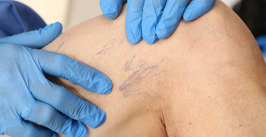
- The formation of spider veins is a network of dilated small veins.
- Well-defined pattern of clogged veins protruding under the skin.
- The formation of points of compression of blood vessels - varicose veins in the form of easily distinguishable tubercles on the legs.
- A change in normal skin color, cyanosis, blackness occurs, the upper layer (dermis) acquires a loose structure.
- Pain, heaviness, stretching and tired legs, decreased mobility, difficulty walking.
- With varicose veins of the lower extremities, the formation of soft tissue swellings is possible.
Ignoring timely treatment leads to serious and dangerous consequences if a person can be cured only through immediate surgical intervention.
Disease classification
According to ICD 10, varicose veins are divided into a disease with ulcer, with inflammation, with ulcer and inflammation, if these signs are absent. According to the 1994 international classification of chronic venous diseases, varicose veins are classified into:
- Intradermal, segmental. No pathological venous discharge is observed.
- Segmental with reversed blood flow, occurs through the superficial and perforating veins.
- Distributed with reverse blood flow through superficial and perforating veins.
- Varicose veins with reverse blood flow through the deep veins.
It is customary to classify the disease according to additional signs of the clinical picture:
- There are no symptoms on examination or palpation.
- The reticular veins are expressed.
- There are varicose veins.
- There is swelling of the soft tissues.
- Violation of normal skin color.
- Revealed lipodermatosclerosis.
- There is a healed ulcer.
- An active ulcer was shown.
Symptoms are absent or subjective (patient's feelings). In addition, varicose veins are classified on the basis of reasons: congenital, primary, secondary, with an unknown factor that triggered the development of the disease.
Diagnosis of varicose veins
The predominant method of detecting varicose veins is visual examination and palpation of the patient. In order to carefully determine the severity of the disease and choose the right treatment, the phlebologist, when examining the anamnesis and palpation, prescribes:
- A complete blood count is the main standard for determining red blood cell counts and hemoglobin levels. Based on the blood coagulation, conclusions are drawn about the degree of development of the disease and the predisposition to thrombosis.
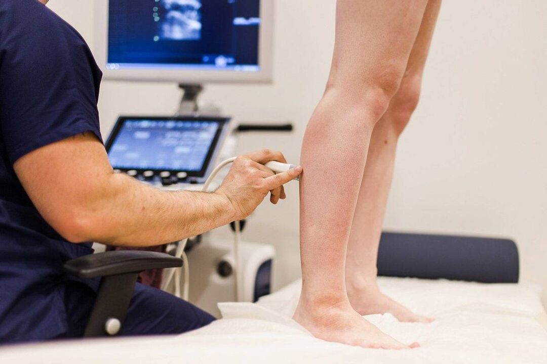
- Doppler ultrasound. The procedure consists in ultrasound diagnosis of the speed and direction of blood particles. In this way you can determine which direction the blood is flowing in and whether it is at a sufficient speed.
- Ultrasound agnioscanning. It is used to visually examine the vessel walls, their structure, direction and speed of blood flow in real time on the monitor of the ultrasound device.
- Plethysmography. The diagnosis is based on the detection of the electrical resistance of the leg tissue. With proper circulation, the parameter should indicate the normal standard.
- Rheovasographic diagnostics. Based on the determination of the tissue blood filling index. The rheographic index helps determine the stage of varicose veins - compensation, subcompensation or decompensation.
The history of the disease and its examination, which are given comprehensive diagnostic data, allow the doctor to choose the method of treatment.
Conservative drug therapy
This method of treatment uses special drugs that have a positive effect on the course of the disease. Conservative treatment of varicose veins is effective at the initial stage and is used as an additional method of treatment in the formation of lumps, ulcers and eczema.
The main groups of prescribed drugs are:
- Phlebotonics and Phleboprotectors. Venotonic drugs are the standard, which implies conservative treatment. Promote the restoration of the structure of the vascular walls, strengthen and tighten blood vessels.
- Effective blood thinning agent. They help improve the quality composition, the blood flows faster through the veins, reduce the risk of blood clots, restore normal blood circulation and relieve pain.
- NSAIDs (anti-inflammatory drugs). Eliminate pain, prevent cramps, effectively relieve inflammation and swelling.
Conservative treatment will help in timely referral to a phlebologist, at the initial stage it is possible to influence the composition of the blood and the condition of the vascular walls. With complex forms of the disease, drastic measures are required.
Laser therapy
Laser therapy is considered a gentle and least traumatic method when varicose veins of the legs have to be treated, classified according to ICD 10 code 183. The idea of the method is to use a laser beam that actively affects the vessel walls and promotes their adhesion. An LED attached to a laser device is inserted into the vein by piercing the skin. The beam is selective and has no influence on adjacent healthy tissue. Main advantages of laser therapy in the treatment of varicose veins:
- Quick positive effect.
- No pain and injuries.
- Stable result, long-term remission.
- Restoration of normal blood circulation.
Contraindications for use are thick or too thin vessel walls, large venous lumens, pregnancy, oncology and other serious comorbidities.
Sclerotherapy of varicose veins
The method is based on the introduction of special liquid or foamy preparations - sclerosants - into the vessels affected by varicose veins. They replace endothelial cells with fiber tissue. Needles, syringes, and sclerosants are used to perform sclerotherapy.
The treatment technique consists of the following steps:
- piercing a pathological vein;
- Pumping out (removing) all of the blood from the vessel;
- Administration of a sclerosant preparation;
- the application of a suitable bandage or knitted compression.
This method gives a lasting result. The procedure is painless; the fusion of vascular tissue with varicose veins is an alternative to surgery.
Perform an operation
The most painful and traumatic way to treat varicose veins is through surgery. Indications for its implementation are extensive vascular lesions, the presence of varicose veins, dangerous consequences of the disease, for example, acute thrombophlebitis.
Phlebectomy is performed under local anesthesia, the pathological vein is ligated, the required number of incisions are made to remove it, and are removed. The operation is recognized as an effective treatment method, showing the result in eighty percent of the cases. However, phlebectomy has a number of side effects: wound complications, trauma to the lymph nodes, in extreme cases with damage to deep nerves, immobilization and disability can occur.
To avoid dangerous complications of varicose veins, which are manifested by: lumps, ulcers, bleeding, phlebothrombosis, pulmonary embolism and other serious consequences, you should consult a doctor at the initial stage of varicose veins!